By Nicholas Theodore, M.D.
If a person’s spine is unstable because of injury, degenerative disease or another cause, he or she may need spinal stabilization surgery to correct the problem. During this procedure, surgeons typically take multiple X-rays to pinpoint where to place screws to stabilize the spine.
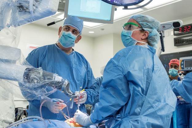
I was convinced there was a better way than using numerous X-rays, so I co-invented an image-guided robot that uses a computed tomography scan taken before or during surgery to precisely plan and insert the screws. Now, with a push of a button, a robot guides surgeons in placing spinal screws with submillimeter accuracy.
The idea came from thinking about how to protect surgical team members as they perform spinal stabilization procedures day to day. To place the screws, patients used to get 10 to 20 X-ray scans, which we consider a safe threshold of radiation exposure. But for the surgical team, the radiation exposure becomes excessive over time—although we do wear lead protection—putting us at risk for cancer.
I wondered if I could take the X-rays either before or during surgery without being in the room. Could I put those images into a system where I can navigate the spinal column without having to shoot additional X-rays? By taking a scan before or during surgery, the surgical team steps behind a radiation shield and suffers no exposure. The robot has essentially dropped our radiation exposure to zero, while also collapsing surgical time.
With the robot, we plan the trajectory. Before we put the screw in, I draw a line on the screen and place a virtual screw in the bone. When I put the real screw in, I know precisely how long and how wide the screw should be based on the patient’s anatomy.
In typical image-guided surgery, the surgeon watches the screen. However, since the robot is locked on my trajectory as I’m putting the screw in, I can look at the patient and his anatomy with my own eyes. It allows us to elevate and perfect this concept of minimally invasive surgery.
To give an example, I performed a spinal surgery where it took the robot five minutes to help me place six screws through a minimally invasive incision. I couldn’t do that with the old X-ray method. Patients now can go home in one or two days instead of four or five because of reduced time under anesthesia and less tissue disruption, so healing can happen faster.
I’m very proud that The Johns Hopkins Hospital was the first of more than 60 health care systems worldwide now using this innovation. I’m also very excited to see where this technology will lead. Right now, we’re looking at using the robot to target the brain to do a tumor biopsy or put a brain electrode in someone with Parkinson’s disease. The reality is we can use this technology to target any place on the body. I truly believe his combination of imaging, surgery and robotics will fundamentally change the way we do surgery.
A version of this story first appeared in Leader, a magazine that connects the leaders of the world with Johns Hopkins Medicine, a global leader in medicine.
Nicholas Theodore, M.D., director of the Johns Hopkins Neurosurgical Spine Center, is an internationally recognized expert in brain and spinal cord injury, minimally invasive spine surgeries and robotics. As an award-winning teacher and researcher, Dr. Theodore has written or co-authored 50 book chapters and more than 200 peer-reviewed journal articles, and he is co-holder of more than 20 patents for medical devices and procedures. His research focuses on trauma, spinal cord injuries, robotics, and developing an understanding of the genetic and molecular basis of spinal diseases.
